An estimated one in 11 people is diagnosed with post-traumatic stress disorder, or PTSD. People with PTSD struggle with intense anxiety that lingers long after the traumatic event, which can make day-to-day life extremely difficult. But now, a new study sheds light on brain characteristics of individuals with PTSD, paving the way for innovative treatments in the future. Science Daily reports that the study, a collaboration between the San Francisco Veterans Affairs Medical Center and researchers at UC Berkeley, used a number of tests – including the popular open field test – to explore anxiety behavior in rats with increased myelin levels, a protective substance that wraps around axons of brain neurons. Read on to find out more about the connections between myelin, anxiety, and PTSD.
What Is Myelin?
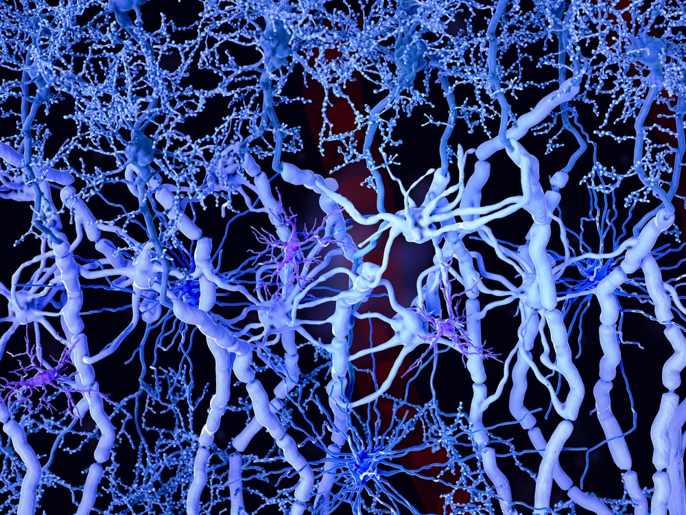
Myelin, sometimes referred to as the myelin sheath, consists of a layer of fatty substances and proteins. That layer wraps around the axons of neurons in the brain to form, as Science Daily puts it, “the insulation around the brain’s wiring.” With that, myelin helps to facilitate effective communication between different areas of the brain. Myelin is whitish in color, and made of bundles of long axons – hence why those areas are called “white matter.” Far less myelin exists in other areas that are mainly formed by groups of cell bodies, which are collectively known as “gray matter.” However, researchers at the San Francisco Veterans Affairs Medical Center recently found unexpected quantities of gray matter myelin in veterans with PTSD. This raised a question for the researchers: How did the presence of myelin link back to the veterans’ PTSD symptoms?
How Is Myelin Related to Anxiety and PTSD?
The veterans studied at the San Francisco Veterans Affairs Medical Center showed increased myelin, particularly in areas of the brain associated with emotions and memory. The researchers realized that myelination in these “memory zones” could, in fact, be a side effect of the veterans’ trauma, further contributing to their lasting PTSD symptoms. These findings also suggest that specific PTSD symptoms could be related to which areas of the brain have been myelinated. To test that theory, the San Francisco Veterans Affairs researchers collaborated with colleagues at UC Berkeley to explore the presence of myelin in rat models. The UC Berkeley team employed a number of tests, including a popular behavioral assay known as the open field test.
Using the Open Field Test
First, the UC Berkeley researchers had to observe their rat subjects during periods of intense stress. To accomplish this, the researchers exposed rats to an acute, severe stressor and afterwards employed a number of behavioral tests, including the popular open field test. During the open field test, researchers assessed the general activity levels and anxiety-type behaviors of rats left to explore an open space. By the end of the study, the Berkeley researchers had subjected the rats to a whole battery of tests to evaluate stress and anxiety levels. In addition to the open field test, the tests included several common behavioral assays for rodent models:
- An elevated plus maze, during which rats are placed at the center of a maze and assessed based on their willingness to explore, testing rats for anxiety
- A light-dark box, where the rodents are observed to see if they decide to explore novel areas or if they’d rather avoid brightly lit, open spaces, testing them for anxiety
- An acoustic startle response test, during which rats are observed after being exposed to a surprising sound stimulus that tests for startle response
- A fear conditioning test, during which rats are exposed to a neutral conditional stimulus (a loud sound) in combination with an unpleasant unconditional stimulus (an electric stimulus) testing for emotional learning and memory
Conclusions from the Open Field Test
After all of the tests were concluded, the UC Berkeley researchers were able to evaluate myelin in the rats. As expected, they saw increased myelin levels in the rats’ gray matter. Ultimately, the researchers concluded that stress produces more of the brain’s glial cells, called oligodendrocytes, that are the main producers of myelin. Those cells then wrap around cell bodies of neurons, which are located in the gray matter. The increased myelin could disrupt cellular communication and make the subject hyper-responsive to fear. With this in mind, researchers can now explore myelin manipulation with the goal of advancing PTSD treatment.
_____
Researchers are still unsure of how to shape myelin content in the human brain. However, future research could result in treatments to help reverse myelin buildup in gray matter – thus reversing the effects of PTSD.
Scantox is a part of Scantox, a GLP/GCP-compliant contract research organization (CRO) delivering the highest grade of Discovery, Regulatory Toxicology and CMC/Analytical services since 1977. Scantox focuses on preclinical studies related to central nervous system (CNS) diseases, rare diseases, and mental disorders. With highly predictive disease models available on site and unparalleled preclinical experience, Scantox can handle most CNS drug development needs for biopharmaceutical companies of all sizes. For more information about Scantox, visit www.scantox.com.