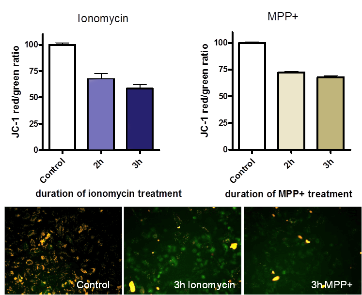
Differentiated SH-SY5Y human neuroblastoma cells were lesioned with the Calcium ionophore ionomycin or with MPP+. Lesions were examined with JC-1 for mitochondrial membrane depolarization and with the MTT assay for cell viability in 96-well plates. JC-1 labelling was determined in a 96-well fluorescence plate reader with the appropriate filter settings. The ratio of red/green fluorescence in control cells was set to 100%. Compounds can be examined for their ability to counteract mitochondrial membrane depolarization induced by selected lesion agents.

Figure: Effect of ionomycin and MPP+ on mitochondrial membrane depolarization by JC-1 labeling. Images indicate control cells and cells lesioned with ionomycin or MPP+ for 3 hours. In intact mitochondria, JC-1 forms red fluorescent aggregates, whereas green fluorescent monomers indicate mitochondria with depolarized membrane potential.
Remark: These assays can also be customized for primary neurons.
