This approach allows you to evaluate the effect of your compound in an intact neural system. By maintaining the intact 3D structure and cell-cell interactions of the postnatal brain, this system comes closest to in vivo models.

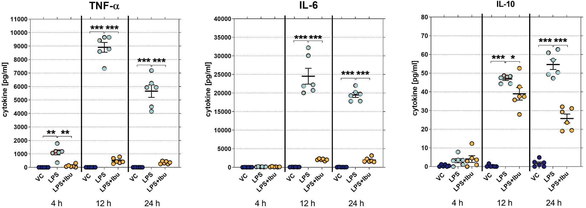
Cytokine release by mouse hippocampal slices after LPS stimulation over time. Data are displayed as aligned dot blots with group means (n=6 per group). Mean ± SEM. Two-way ANOVA followed by Bonferroni`s multiple comparison post hoc test compared to LPS group. *p<0.05, **p<0.01, ***p<0.001.

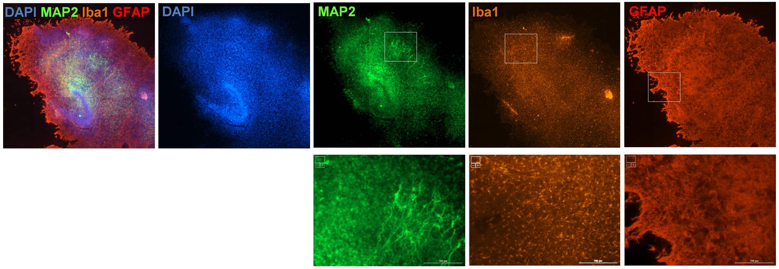
Representative images of mouse hippocampal slices fixed after 24 h of LPS stimulation. Slices were immunohistochemically labeled using the astrocytic marker GFAP (red), the microglia marker Iba1 (orange) and the neuronal marker MAP2 (green) as well as nuclear stain DAPI (blue). Scale bar: 200 µm.
