Solution 1
PD involves the degeneration of nigrostriatal dopaminergic (DA) neurons. This pathology can be modeled in experimental animals by the administration of MPTP (1-metyl-4-phenyl-1, 2, 3, 6-tetrahydropyridine), a neurotoxin which is converted in the brain into MPP+ (1-methyl-4-phenylpyridinium) by the enzyme MAO-B a toxin which selectively destroys DA neurons and causes anatomical, behavioral, and biochemical changes similar to those seen in Parkinson’s disease.
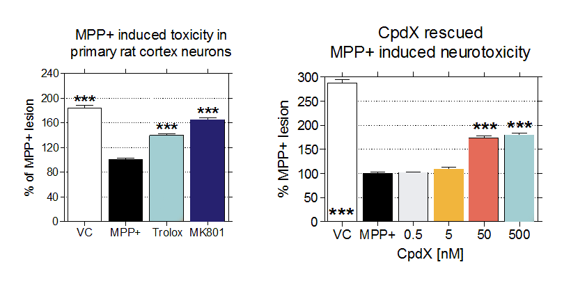
In vitro Parkinsonism can be modeled by the administration of MPP+ to different cell culture systems. Scantox provides several cell culture models for MPP+ induced Parkinsonism, as primary cortical neurons (Figure), primary rat TH neurons, SH-SY5Y and many more.

Figure 1: Effects of known compounds as Trolox and MK-801 (left) and Compound X (right) on MPP+ induced toxicity in primary rat cortex neurons.
Solution 2
A53T α-synuclein preformed fibrils PD additionally involves accumulation of α-synuclein protein in different brain regions. Recombinant human α-synuclein (α-syn) fibrils can be used to evaluate seeding properties and induce toxicity on primary cortical neurons in vitro.
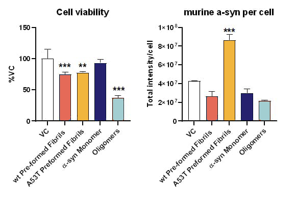
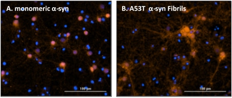
While monomeric human α-syn has no impact on cell viability, preformed wild type and A53T human α-syn fibrils have toxic effects on primary cortical neurons. This toxic effect is exceeded by oligomeric isoforms (Fig.1A). Only A53T α-syn fibrils of the tested isotypes show seeding properties in cortical neurons (Fig.1B).

Figure 2. In vitro assessment of α-syn preformed fibrils on mouse primary neurons. Toxicity (A) as well as seeding (B) properties of different preformed recombinant human α-syn species (Stressmarq) on mouse primary cortical neurons. (A) Neurons treated with α-syn species and assessed for cell viability by MTT assay. (B) Neurons treated with α-syn species and immunocytochemically analyzed for murine α-syn. Mean+SD. One-way ANOVA with Bonferroni post hoc test (vs vehicle control: VC). ** p<0.01, *** p<0.001.

Figure 3. Representative images of endogenous murine α-syn accumulation after seeding with different preformed recombinant human α-syn species (Stressmarq). Neurons were treated with (A) monomeric and (B) A53T preformed fibrils and after incubation immunocytochemically stained for murine α-syn. Nuclear stain DAPI = blue; murine α-syn = red; scale bar 100 µM.
