TDP-43 Induced ALS Mouse Model
The TAR DNA binding protein (TARDBP or TDP-43) is shown to play a crucial role in a growing set of neurodegenerative diseases. While strongly related to sporadic and familial forms of amyotrophic lateral sclerosis (ALS), intraneuronal TDP-43 accumulation and aggregation is also related to frontotemporal lobar degeneration (FTLD-TDP or formerly FTLD-U).
To induce ALS in C57BL/6 mice, animals are injected into the motor cortex with adeno-associated viral (AAV) particles of serotype 9 that express human TDP-43 (AAV9-GFP-hTDP-43). Control animals are injected with AAV9-GFP. Analyzing the animals which were induced at 3 months of age and followed over the next 6 months, the AAV9-GFP-hTDP-43 injected animals show:
- Early muscle weakness
- Learning deficits
- hTDP-43 expression in cortex
- Increased activated microglia
- Increased ubiquitination
- Neuronal loss
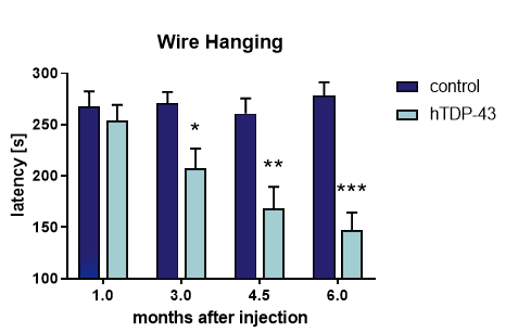
Muscle weakness can be observed already 3 months after AAV9-GFP-hTDP-43 injection (Figure 1).

Figure 1: Wire hanging in AAV9-GFP-hTDP-43 and control mice. Latency to hang on the wire up to 300 s was measured. Two-Way-ANOVA with Bonferroni post hoc test. Mean + SEM. Control: n = 16, hTDP-43: n = 20. * p < 0.05, ** p < 0.01, *** p < 0.001.
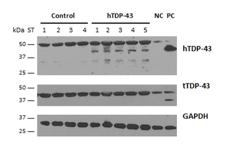
Six months after AAV9-GFP-hTDP-43 injection, mice express hTDP-43 protein in the cortex (Figure 2).

Figure 2: Expression of human and total TDP-43 in the cortex of control and hTDP-43 mice. Western blot analysis of cortex samples of AAV9-GFP-hTDP-43 and control mice 6 months after injection, homogenized in RIPA buffer and with GAPDH as loading control. PC: positive control = hippocampal RIPA sample of transgenic TDP-43 mouse. NC: negative control = untreated wild type mouse.
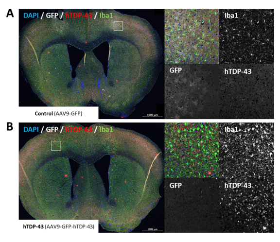
hTDP-43 expression and increased levels of activated microglia are measurable in the brain of AAV9-GFP-hTDP-43-injected mice after 6 months (Figure 3).

Figure 3: Immunolabeling of GFP (white), human TDP-43 (red), Iba1 (microglia marker, green) and nuclei (DAPI, blue) in the brain of control mice (A) and AAV9-GFP-hTDP-43 injected (B). Coronally cut brain samples at 0.62 mm. White box: Area of injection magnified on the right. The virus was injected bilaterally into the motor cortex region M1 of 3 months old mice. Samples were collected 6 months after injection.
Scantox offers a custom-tailored study design for the induced ALS model, and we are flexible to accommodate your special interest. We are also happy to advise you and propose study designs. Virally induced hTDP-43 mice are a valuable model of slowly progressing ALS. Vehicle-injected wild type mice can serve as control needed for proper study design. This model is therefore a proper model to evaluate the efficacy of potential treatments against ALS and to observe the time dependent effects of such a treatment.
We are happy to evaluate the efficacy of your compound in an induced ALS animal model! The most common readouts include:
- Motor deficits
- Memory performance
- hTDP-43 expression in cortex
- Ubiquitination
- Neuroinflammation
- Neuronal loss
You might also be interested in these related topics:
- ALS Transgenic Mouse Models
- TDP-43 Transgenic Mouse Models
- Lipopolysaccharide (LPS) Induced Neuroinflammation
